Leishmaniasis is a disease caused by protozoa of the genus Leishmania, transmitted by mosquitoes.

Content
Leishmania
This is a genus of parasitic single-celled animals. In the body of a warm-blooded animal, they are in the stage of a flagellated intracellular form. In the body of the vector and on nutrient media they exist in a flagellar form.
The life cycle of Leishmania occurs with a change of hosts - a vertebrate animal or a human and a mosquito.
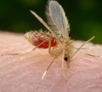
The mosquito (the carrier of the disease) is a small dipterous insect measuring 1.2-3.7 mm. It becomes infected by blood-sucking from an infected vertebrate animal or human. In the intestines of the insect, Leishmania develops, multiplies and within a week turns into infectious forms, which are concentrated in the anterior intestines and proboscis of the mosquito.
With repeated blood sucking, infectious forms (promastigotes) from the mosquito enter the blood of the vertebrate host and are absorbed by immune cells. There they develop and reproduce. Cells overcrowded with parasites are destroyed, the parasites are captured by other cells, where the reproduction process is repeated.
In warm countries, mosquitoes are active all year round. These are crepuscular and nocturnal insects. During 2-3 weeks of life, females feed on blood and lay eggs 2-3 times.
The main natural reservoir of the parasite are rodents and representatives of the canine family.
Leishmaniasis
There are several types of leishmaniasis.
Diseases characteristic of Europe, Asia, Africa:
- Indian visceral (kala-azar),
- Mediterranean-Central Asian,
- children's visceral,
- zoonotic (rural) cutaneous,
- anthroponotic (urban) cutaneous,
- Mexican skin
America has its own types of leishmaniasis:
- Mexican (Chiclero ulcer),
- Peruvian (Uta),
- Guianan (forest yaws),
- Panamanian,
- mucocutaneous (espundia).
Indian visceral leishmaniasis (kala-azar)
 This type of disease is common in India, Bangladesh, Nepal, northeastern China, Kenya, Somalia, Sudan, Uganda, and Ethiopia.
Children aged 5-9 years are most often affected. Those who have recovered from the disease acquire stable and long-lasting immunity. Recurrent diseases are practically not recorded. In patients with AIDS, leishmaniasis becomes malignant and leads to death.
At the site of the bite of an infected mosquito that carries the disease, a tumor forms on the skin. Leishmania, multiplying, penetrates the lymph nodes, then moves to the spleen, bone marrow, liver, lymph nodes of the intestines and other internal organs.
As a result, the functioning of these organs is disrupted and they increase in size. The spleen is most affected, anemia progresses, which is aggravated by damage to the bone marrow.
In some cases, patients develop the so-called postkala-azar cutaneous leishmaniasis - the appearance of nodules and spotty rashes on the skin of the face and other parts of the body that contain leishmania. These formations persist for many years, and the patient continues to remain a source of infection for a long time.
Symptoms of visceral leishmaniasis
The incubation period ranges from 3 weeks to 12 months, in rare cases it can reach up to 2-3 years. The disease begins gradually.
During the development of the disease the following are observed:
- undulating fever,
- enlarged spleen,
- liver enlargement,
- enlarged lymph nodes,
- diarrhea,
- darkening of the skin,
- hemorrhages in the skin and mucous membranes,
- nasal and gastrointestinal bleeding.
On palpation, the liver and spleen are dense and painless.
In HIV-infected people, the disease is malignant and is accompanied by resistance to specific therapeutic drugs.
Diagnostics
The diagnosis is made on the basis of characteristic symptoms and confirmed by laboratory tests.
Leishmania is detected in punctate smears of bone marrow, spleen or liver. Leishmania is found extremely rarely in peripheral blood.
Treatment
For treatment, pentavalent antimony preparations are used - solyussurmin or meglumine antimonate. The course of treatment is 30 days. If the disease relapses, repeat after 14 days.
In severe cases or ineffective treatment with antimonial drugs, second-line drugs are used - amphotericin (for adults) and paromomycin (for adults and children).
Mediterranean-Central Asian leishmaniasis
Another name is infantile leishmaniasis. The disease is common in Mediterranean countries, with isolated cases recorded in Asia, Transcaucasia and Crimea.
Symptoms
The incubation period ranges from 1 month to 1 year.
The main distinguishing signs of the disease are:
- no skin rashes,
- inflammation of the lymph nodes,
- stomach ache,
- coughing attacks.
Often accompanied by bacterial pneumonia. The skin is pale and has an earthy tint.
Acute and subacute forms are rare, mainly in young children. These forms of the disease are severe, require immediate treatment and are characterized by sudden exhaustion of the patient, decreased muscle tone, the appearance of boils, and ulcerative lesions of the oral cavity.
The most common form of the disease is chronic, which is observed mainly in older children, less often in adults. This form is characterized by a milder course, attacks are replaced by long periods of normal well-being. With timely treatment, complete recovery occurs. Even an enlarged liver and spleen quickly shrink to normal size.
A significant number of cases of infection are asymptomatic or in an erased form; spontaneous recovery may occur.
In people with immunodeficiency, especially in HIV-infected people, the disease develops quickly and is very severe, difficult to treat and often leads to the rapid death of the patient.
Treatment is the same as for Indian visceral leishmaniasis.
Zoonotic (rural) cutaneous leishmaniasis
 Found in the countries of the Middle East, North and Central Africa, Central Asia, Kazakhstan, Mongolia, Transcaucasia, Iran, Afghanistan.
Humans are not the source of infection. The main natural reservoir of infection is rodents.
After suffering the disease, stable lifelong immunity to all forms of cutaneous leishmaniasis is developed. For example, in the south of Turkmenistan, the majority of the local population becomes ill in childhood.
Symptoms of rural cutaneous leishmaniasis
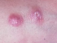
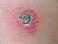
- The disease begins with the appearance at the site of a mosquito bite of a tubercle 2-4 mm in size, surrounded by a rim of inflamed skin.
- On the second day it increases to 8-10, sometimes up to 15 mm in diameter.
- At the same time, the inflammatory swelling of the skin around it increases.
- After 1-2 weeks, an ulcer with steep, steep edges with a diameter of 2-4 mm appears in the center.
- The bottom of the ulcer is uneven, covered with a yellowish-gray or yellowish-green coating.
- Ulcers can be multiple with multiple bites.
- The ulcers are not painful, but pain occurs during dressings or accidental injuries to the ulcers (blows, pressure).
- The appearance of severe pain in ulcers indicates the addition of a secondary bacterial pathogenic infection.
- At 2-3 months, the ulcers are cleared of pus.
- At 2-6 months their scarring begins.
Treatment
If the disease does not really bother the patient and is not complicated by infection of the ulcers, then treatment is not carried out and the disease is allowed to develop naturally.
The drugs are used when the disease is complicated by inflammation of the lymph nodes.
Prevention
The most effective method of preventing the disease is vaccination.
Anthropotic (urban) cutaneous leishmaniasis
 Found in Europe, Asia, America, Africa, Central Asia and Transcaucasia.
The source of infection is a sick person, an additional reservoir is dogs.
The disease occurs throughout the year. Among the local population, children are mostly affected; among visitors, people of all ages are affected.
Symptoms
The incubation period ranges from 2-4 months to 1-2 years, sometimes lasting up to 4-5 years.
After this period, at the site of the bite of infected mosquitoes (usually on the face and upper limbs), barely noticeable single, less often multiple, tubercles (leishmaniomas) with a diameter of 2-3 mm with a smooth, shiny surface appear.
They slowly increase and after 3-4 months reach 5-10 mm in diameter, acquiring a reddish-brown color with a bluish tint.
After several months, the tubercles may resolve. However, this course of the disease is rare.
As a rule, a scale forms on the surface of the tubercle, which then turns into a yellowish-brownish crust tightly attached to the tubercle.
After the crust falls off or is forcibly removed, a bleeding erosion or ulcer is discovered. For a long time, the ulcer is covered with a dense crust.
After 2-4 months, scarring of the ulcers gradually begins, which ends on average a year after the appearance of the tubercle. In some cases, the disease drags on for 2 years or more.
Complications
In 10% of patients, sluggish chronic tuberculoid recurrent cutaneous leishmaniasis develops. Its symptoms are similar to tuberculous lupus and can last for decades. Presumably the cause of this complication is immunodeficiency.
Another complication is pyodermatitis, which develops due to the addition of a secondary infection.
Diagnostics
First of all, it is determined whether the victim was in areas where this parasite is spread.
Tissue taken from the edge of the lesion is analyzed.
Treatment
If the disease does not really bother the patient, then treatment is not carried out. In the early stages of the formation of tubercles, they can be treated with specific drugs or ointments containing chlorpromazine (2%), paromomycin (15%) or clotrimazole (1%).
Treatment for tuberculoid leishmaniasis is the same as for visceral leishmaniasis, but it is difficult to treat. Repeated courses are often necessary in combination with immunostimulants, vitamins and restorative drugs.
H2Mexican cutaneous leishmaniasis
This type of disease is common in Latin America, Mexico, Peru, and the USA.
Immunity after an illness is unstable.
Symptoms
The incubation period ranges from 2-3 weeks to 1-3 months.
The disease usually occurs in a relatively mild form; one leishmanioma forms on open, accessible parts of the body. It heals without complications after a few months.
However, when leishmanioma occurs on the auricle (40% of cases), the disease takes a long, chronic course and leads to deformation of the auricle.
There are isolated cases of the formation of deep ulcers and destruction of nasal cartilage.
Diagnosis and treatment are the same as for European cutaneous leishmaniasis (urban leishmaniasis).