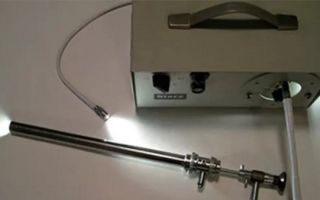
A proctological examination is an external examination of the perianal area, examination of the rectum with a finger, anoscopy, sigmoidoscopy or sigmoidoscopy with a flexible apparatus.
Content
What can be determined by digital examination?
The examination by the proctologist is carried out in the knee-elbow position.
Examination of the rectum with a finger is important in recognizing hemorrhoids, papillitis, anal fissures , rectal fistulas (paraproctitis), strictures and cryptogenic ulcers, rectal prolapse .
With a digital examination, the doctor can identify tumors. This is especially important when small tumors are located in the distal rectum, as they can be easily missed during recto- and colonoscopy if attention is not specifically focused on this area.
During the examination, pay attention to the tone of the sphincter. If the sphincter is insufficient, the patient may experience itching, the skin around the anus is macerated (swollen tissue).
Increased tone and narrowing of the anus are also detected.
Anoscopy
This type of examination is carried out using special mirrors - anoscopes. There are many types of mirrors, including those with lattice and solid doors. Practice shows that mirrors with solid doors are less traumatic.
An anoscope lubricated with Vaseline oil is inserted into the anus with the valves closed, and then they begin to carefully open them.
The use of rectal mirrors allows you to see the mucous membrane of the lower ampullary rectum and anal canal.
- This method is of particular importance in the following cases. It allows you to distinguish true polyps from hypertrophied papillae. The latter represent an elevation of the intestinal mucosa as a result of chronic irritation of the intestine due to anal fissures, proctitis and other chronic diseases. Often hypertrophied papillae acquire a polypoid shape. Incorrect diagnosis leads to unnecessary surgery.
- Makes it possible to distinguish between a fibrous polyp and a thrombosed internal hemorrhoid.
Sigmoidoscopy
This method is intended for a more in-depth examination of the rectum and sigmoid colon.
Sigmoidoscopy should be preceded by a digital examination. It has not only diagnostic value, but also prepares the anus for the introduction of a sigmoidoscope.
The procedure is performed in a knee-shoulder position, in which the patient leans on the table with his left shoulder. The back should be arched. This position straightens the rectosigmoid colon and facilitates examination, while requiring little or no air to dilate the colon.
If a person is weakened and cannot take this position, then the examination is carried out in a position lying on his side, but the examination is much more difficult.
Many patients are concerned about how painful this procedure is. If the procedure is performed by an experienced doctor, then sigmoidoscopy will not hurt, only some discomfort is possible. And to reduce spasms, it is enough to take several slow and deep breaths during the procedure.
The duration of the examination is usually 10-20 minutes.
Indications for retoromanoscopy
Sigmoidoscopy helps to identify the problem even before the first symptoms appear. Therefore, doctors recommend being examined from time to time for preventive purposes.
In addition, indications for sigmoidoscopy are such proctological problems as:
- haemorrhoids;
- inflammation of the rectum ( paraproctitis , etc.);
- rectal bleeding, mucus, pus in the stool;
- long-term constipation;
- diarrhea (diarrhea);
- weight loss;
- suspicion of a malignant tumor of the prostate gland in men, sigmoid or rectum.
During the procedure, an analysis can be taken for histological examination to determine the nature of the possible neoplasm.
Preparation for sigmoidoscopy
Careful preparation for the procedure is a necessary condition for its implementation. The presence of feces even in some parts of the intestine can lead to the fact that a polyp, for example, will not be detected.
The following are recommendations from ONKlinik on preparing for the procedure.
- Before the initial examination, it is necessary, no later than 3-4 hours before the appointment, to cleanse the intestines of feces.
- An exception is for patients who experience severe pain in the anal canal: they can come for an examination without preparation.
- The patient can choose one of the proposed preparation schemes, which will be more comfortable and accessible for him.
Preparation option using cleansing enemas
On the eve of the study, at 20.00, it is necessary to perform a cleansing enema using 1-1.5 liters of boiled water cooled to room temperature.
- Enemas are performed in the “lying on the left side” position; after administration, you must refrain from eating. The exception is for patients suffering from diabetes (they are advised to have a light dinner).
- On the day of the examination, in the morning (at 6.00), a cleansing enema is performed according to the same scheme.
After morning enemas, you must refrain from breakfast. Patients suffering from diabetes must take breakfast with them.
Preparation option using microenemas “MICROLAX”
"MICROLAX" is a laxative drug with combined action. It is produced in the form of microenemas, which are polyethylene tubes with a tip and a removable seal, each tube containing 5 ml.
How to use?
- It is necessary to remove the protective seal on the tube and lightly press on it to lubricate the tip of the enema and facilitate the insertion process. Next, you need to insert the microenema into the entire length of the rectum and squeeze out the entire contents of the tube. Then slowly remove the tip while continuing to gently squeeze the tube.
- For complete emptying, it is enough to insert two or three tubes at intervals of 5-10 minutes. The laxative effect is observed approximately 5-15 minutes after administration of the drug. This set of procedures should be carried out no later than 3 hours and no earlier than 6 hours before the expected time of examination.
The drug can be used during pregnancy and lactation (breastfeeding).
Preparation option using the drug “ENEMA CLEAN”
“ENEMA CLEAN” is a saline laxative, the effect of which is due to water retention in the intestinal lumen as a result of osmotic processes. The accumulated liquid enhances peristalsis and helps cleanse the intestines, and the laxative effect occurs within 5-7 minutes after administration of the drug.
How to use?
- Before administration, you must take a supine position: lie on your left side, bend your right leg at the knee. Then remove the protective cap and carefully insert the tip into the anus, after lubricating it with a small amount of the drug.
- After introducing the contents, you must press the bottle with slight force in the direction of the navel. After the contents of the bottle have been squeezed out, the tip must be removed. In this case, it is not necessary to squeeze out the entire drug, since the bottle contains an excess of about 15 ml of medicine.
It is recommended to maintain the body position adopted when administering the drug until the onset of a laxative effect and the urge to defecate.
What is the difference between sigmoidoscopy and colonoscopy?
The two procedures differ in the depth of examination of the intestines.
Sigmoidoscopy penetrates to the sigmoid colon. While a colonoscopy can examine the entire large intestine and part of the small intestine. Sigmoidoscopy is less painful, is usually done without anesthesia and does not require such careful preparation of the patient for the procedure as colonoscopy.


