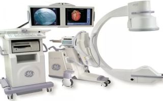
Radiography is a method for diagnosing diseases of organs and tissues, based on the special property of X-rays to penetrate dense opaque media and be absorbed by them to an unequal extent, depending on their chemical composition and physical properties. X-ray examination is still the main one in the diagnosis of intestinal diseases.
Content
Types of X-ray examination
Methods for examining the intestines using X-rays are divided into
- non-contrasting and
- carried out using radiopaque agents.
Fluoroscopy and abdominal radiography are non-contrast methods.
They allow you to detect
- free gas in the abdominal cavity due to perforation of the intestinal wall,
- foreign bodies,
- pathological accumulations of fluid and gas in the intestine due to its obstruction, etc.
X-ray of the small intestine
This study is recommended for all patients with small intestinal disease. It is usually carried out using a contrast agent. The intestine is filled with a suspension of barium sulfate.
The substance is taken orally and after 10-15 minutes an image of the first loops of the jejunum appears, and after 1.5-2 hours all parts of the small intestine are visible on the screen.
In order to speed up the filling of the small intestine with a radiopaque substance (if the function of intestinal motility is not examined), the barium mixture is cooled to 4-5 degrees, in addition, various drugs are administered that stimulate intestinal motor function.
The examination of the small intestine is carried out in both horizontal and vertical positions of the patient.
In some cases, when uniform tight filling of the intestine and its double contrasting are necessary (especially if diverticula of the jejunum and ileum are suspected), the suspension is administered through a probe previously passed through the mouth into the small intestine.
Filling of the intestine is carried out under fluoroscopy control; images are taken in different positions of the patient.
What does an x-ray of the small intestine show?
X-ray examination of the small intestine reveals
- anomalies and malformations,
- tone,
- peristalsis,
- speed and nature of filling,
- intestinal hypersecretion,
- assess the state of the relief of the mucous membrane,
- degree of damage,
- extent of changes.
Study of anomalies and malformations of the small intestine
Anomalies in the development of the duodenum usually manifest themselves in childhood.
X-ray examination allows us to identify pathologies such as
- artresia,
- internal membranes,
- extraintestinal stenosis,
- diverticula,
- elongation,
- excessive mobility.
Most often in clinical practice, diverticula of the duodenum are encountered, which can be single or multiple and reach a diameter of 4 cm.
A mobile duodenum is characterized on X-ray images by elongation, sagging and widening of the lumen, excessive mobility and inflammation.
In the case of the reverse position of the intestine, its horizontal part is expanded, emptying is slowed down.
When part of the intestine is doubled, it is radiologically detected in the form of a tube located parallel to the main intestine.
Study of functional disorders of the duodenum
- Among functional diseases, bulbo- and duodenostasis are often encountered, which are manifested on X-ray examination by a slow passage of barium through the intestine, the appearance of liquid and gases in the lumen.
- Impaired motor function (hypermotor and hypomotor dyskinesias) is manifested by uneven filling of the small intestine with barium sulfate, relaxation of certain areas (regional hypotension) or spasms.
- In cases of severe illness, horizontal levels of liquid and gas are detected in individual intestinal loops. Simultaneously with dyskinesia, acceleration (less than 1 hour) or slowdown (4-6 hours or more) of the passage of suspension through the small intestine is observed.
- Changes in the relief of the mucous membrane are noticeable visually, and are also characterized by uneven accumulations of barium between the altered folds of the mucous membrane after bowel movement.
Study of structural changes in the small intestine
This group of diseases includes:
- tumors
- lymphogranulomatosis,
- intestinal lymphomas,
- lymphomas of the lymph glands of the abdominal cavity,
- Crohn's disease,
- tuberculosis,
- gluten enteropathy (celiac disease),
- Whipple's disease.
If these diseases are suspected, to improve the quality of the study, the barium mixture is also injected directly into the small intestine through a probe (into the distal part of the duodenum or into the initial part of the jejunum).
In some cases, medications cause artificial hypotension of the intestine and an X-ray examination is performed.
Severe damage to the upper small intestine and absence of changes in the ileum in most cases is a sign of celiac disease (gluten intolerance).
X-ray of the large intestine
Filling the colon with a barium suspension through the mouth allows you to evaluate
- motor-evacuation function of the intestine,
- intestine shape,
- position,
- clearance size,
- displacement,
- haustration.
When is it necessary to take an x-ray of the colon?
A transoral examination of the colon is done for the following symptoms:
- long-term persistent constipation ,
- prolonged persistent diarrhea ,
- suspicion of pathology of the ileocecal region,
- suspicion of appendicitis ,
- suspected Crohn's disease.
The main radiological method for examining the relief of the colon is irrigoscopy .
Anomalies and malformations of the colon on x-ray
The examination reveals the following malformations:
- congenital dilatation (megacolon) of the colon,
- lengthening (dolichocolon) of the colon,
- congenital diverticula (recognized both by contrast with an enema and by examination of the intestine 24 hours after ingestion of barium suspension),
- mobile cecum (manifested by pain in the right iliac region),
- Hilayditi syndrome, the location of the right flexure of the colon between the diaphragm and the liver (manifested by vague pain in the upper abdomen, increasing during the day and decreasing at night, bloating and constipation).
Research of functional disorders
To establish violations of the motor function of the colon, x-rays are taken 24, 48 and 72 hours after ingestion of barium suspension.
The disadvantages of this method are the possible inadequate reaction of the intestines to the suspension. In addition, in this case, tumors are not detected.
However, with functional constipation, a decrease in motor function and an increase (spastic constipation) or decrease (atonic constipation) in the tone of the entire intestine or its segments are detected.
Barium suspension is retained in the intestine during constipation and is detected after 72 hours or more.
To identify diseases manifested by structural changes in the colon, irrigoscopy is used.
Contraindications
It is not recommended to conduct such a study if the patient is in a generally serious condition or with acute mechanical intestinal obstruction.
In addition, the presence of electronic devices in the body (heart pacemaker, hearing aids, etc.) is a contraindication to the study.
Preparing for X-rays
For several days before the procedure, it is necessary to exclude from your diet such foods as brown bread, beans, peas, fresh milk, potatoes and cabbage.
These foods contribute to the formation of gases in the intestines, which, in turn, can lead to difficulties in diagnosis. On the eve of the study, it is necessary to cleanse the intestines with an enema. Correct and thorough preparation for the study is necessary to obtain accurate results and reliable diagnosis of the disease.
Are x-rays harmful?
When conducting an X-ray examination, the body is exposed to light radiation, which does not cause significant harm to health.
New generation systems are characterized by a low radiation dose; in some clinics it is calculated individually for each patient. The equipment allows for individual configuration.
Some believe that it is better not to undergo irrigoscopy for people of reproductive age who plan to have children.
After the examination, the doctor must indicate in the chart what dose of radiation you received. This is necessary to take into account in the case of repeated x-ray examinations within a short time.