A coprogram is a comprehensive analysis that allows you to determine the quality of work of the gastrointestinal tract.
Content
- 1 How is feces formed?
- 2 What can be determined by the appearance of stool?
- 3 Changes in the appearance of stool due to gastrointestinal diseases
- 4 Macroscopic examination
- 5 Microscopic examination
- 6 Stool analysis for dysbacteriosis (dysbiosis)
- 7 Stool analysis for dysbacteriosis - normal values
- 8 Chemical research
- 9 Study of intestinal motor function
- 10 Stool analysis for worms and parasites
- 11 Stool analysis - normal values
- 12 How to collect stool for analysis
How is feces formed?
The food lump (chyme), passing through the gastrointestinal tract, turns into feces in the large intestine.
The breakdown and absorption of substances occurs in the oral cavity, stomach and intestines. The composition of feces can indicate disorders in the digestive system. Therefore, scatological examination is prescribed for the diagnosis of many diseases. Feces consist of digested and undigested food residues (muscle fibers, starch, fiber, fats), bacterial flora of the digestive glands, water, desquamated epithelium (dead intestinal cells). The water content in feces is 70-89%. Normally, the daily amount of feces does not exceed 300 g, and the contents pass through the intestines in 24-72 hours (formed feces without a strong odor).
What can be determined by the appearance of stool?
Disruption of the digestive system or functional abnormalities are reflected in the color, smell, quantity and consistency of stool.
- Insufficiency of gastric digestion : stool is mushy or liquid, the smell is fetid, the reaction is sharply alkaline, a lot of connective tissue, poorly digested intestinal fibers, coarse plant fiber and oxalate crystals, microorganisms are detected microscopically.
- Pancreatic insufficiency : polyfecalation (large amounts of daily stool), creatorrhoea (digestible fiber in stool), steatorrhea (fat in stool) with a predominance of neutral fats, stool has an oily appearance.
- Liver failure : acholic stool (light gray or white clayey), high in fatty acids and soaps.
- Insufficiency of small intestinal digestion : liquid feces, an abundance of organic acids, accelerated passage, residues of all types of food.
- Putrefactive dyspepsia : alkaline reaction of feces, sharp putrid odor, abundance of muscle fibers and connective tissue.
- Fermentation dyspepsia : foamy feces, sharply acidic reaction, large amounts of digested and undigested fiber, intracellular starch grains, drops of fatty acids.
- Lecal (ileocecal) syndrome (observed in case of digestive disorders in the proximal colon): feces are often unformed, golden yellow in color, sour odor, slightly acidic reaction, large amounts of digested fiber and intracellular starch, abundant iodophilic flora, slightly altered muscle tissue fiber and broken down fat.
- Distal colitic syndrome (occurs with inflammatory changes in the distal parts of the colon): unformed feces, containing mucus (sometimes in large quantities), a lot of leukocytes, fiber of the intestinal epithelium. There are no or insignificant amounts of food residues.
- Dyskinetic syndrome (intestinal dyskinesia): fecal fragmentation, fecal fragments are shrouded in mucus, there are no undigested food residues.
Based on the appearance of stool, assumptions can be made about some diseases of the gastrointestinal tract.
Changes in the appearance of stool due to gastrointestinal diseases
| Appearance of stool | Diseases |
|---|---|
| Liquid bright yellow or green | Dysbacteriosis, intestinal infections, gallbladder diseases, Crohn's disease, poisoning |
| Light gray, white, clayey (acholic) | Blockage of the bile ducts (stone or tumor), severe liver dysfunction, hepatitis (especially with dark urine), pancreatitis |
| Red color, blood mixed with feces | Bleeding from the colon or rectum, from hemorrhoids . |
| Black in color with a thin, mushy consistency (melena) | Bleeding from the upper intestine |
| Droplets of blood on the surface of stool | Anal fissure |
| Liquid, with dense lumps, with clotted blood, mucus and pus | Chronic ulcerative colitis |
| Mucus in stool | Inflammatory process in the intestines |
| Mucus mixed with feces | Damage to the small, cecum, ascending and transverse colon |
| Mucus on the surface of stool | Inflammation of the sigmoid and rectum |
| Mucus without feces | Intestinal parasites, prolonged constipation, intestinal obstruction |
| Pus in stool | Ulcerative colitis, intestinal tuberculosis |
| Type of rice congee | Cholera |
| Type of pea soup | Typhoid fever |
| Mucus and streaks of blood with a small amount of feces, pus | Dysentery |
| Type of swamp mud | Salmonellosis |
| Light yellow, plentiful, dough-like | Celiac disease (gluten intolerance) |
| Liquid, foamy, with a sour odor, without mucus | Lactase and sucrase deficiency |
| Hard stool | Impaired intestinal motor function, slow movement of stool through the colon, constipation |
| Sheep feces | Slow movement of stool combined with contractions of the colon |
| Reducing the daily amount of feces | Constipation, peptic ulcer , chronic colitis |
| Increased stool mass (polyfecal matter) | Impaired absorption of the small intestine, increased peristalsis |
| Ribbon shape | Narrowing or prolonged spasm of the sigmoid or rectum |
| Green loose stools in babies | It is normal if the baby is breastfed and the stool does not have a strong unpleasant odor |
Stool examination consists of several stages:
- macroscopic;
- chemical;
- microscopic;
- bacteriological.
Macroscopic examination
Macroscopic examination includes assessment of the amount of feces, its physical properties (consistency and shape, color, smell), as well as visible impurities.
There is no strictly defined amount of daily feces. For example, when plant foods predominate in the diet, its amount increases, and when protein predominates, it decreases.
When using the Schmidt diet, the normal amount of feces is 200-250 grams. In some diseases of the intestines and pancreas, accompanied by impaired absorption processes, a significant increase in the daily amount of feces (polyfecal matter) may be observed.
The consistency of stool (shaped, mushy, watery) and its shape depend on the food consumed. This indicator is usually assessed using the Bristol scale .
Stool color
The color of stool is determined by the presence of the pigment stercobilin; in healthy people it should range from light to dark brown. However, the color can change significantly when taking certain types of food and a number of medications (bismuth and carbolene, as well as blueberries cause black stool, iron - greenish-black, beets - red, etc.).
Changes in stool color are a valuable diagnostic sign for many diseases.
When the flow of bile into the intestines stops, the stool becomes discolored; when bleeding from the upper parts of the digestive tract occurs, black tarry stool is called melena; when bleeding from the colon, the stool may be mixed with unchanged blood and becomes red in color.
With some intestinal infections, stool has pronounced characteristic signs. For example, with cholera, stool resembles rice water, and with typhoid fever, it resembles pea soup.
Stool smell
The smell of feces is caused by organic compounds formed during the breakdown of food proteins. The main components are aromatic substances such as skatole, indole, phenol, hydrogen sulfide and methane.
A change in odor may be due to the following reasons:
- foul-smelling feces are observed during putrefactive processes in the intestines - dyspepsia, tumor decay, etc.,
- sour for fermentative dyspepsia,
- the smell of rancid oil during accelerated evacuation from the large intestine.
Pathological impurities in feces include mucus, blood, pus, helminths, etc.
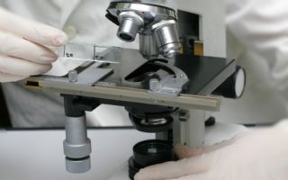
Microscopic examination
This type of stool examination allows you to diagnose functional disorders in the digestive system, inflammatory processes in the digestive tract, other pathologies, as well as protozoa and helminths.
- Normal feces, when examined microscopically, are a detrital, amorphous, fine-grained mass consisting of tiny particles of food debris.
- In healthy people, semi-digested muscle and connective fibers, which are remnants of protein foods, are contained in very small quantities (1-2 fragments of altered muscle fibers in the field of view at low magnification).
- The appearance of a large number of muscle fibers, especially those that retain transverse striations (creatorrea), indicates insufficiency of pancreatic function or a decrease in the secretory function of the stomach.
- Indigestible fiber (coarse parts of plant foods, skins of vegetables and fruits) is not broken down in the intestines and is excreted in the feces. Digestible fiber and starch are completely digested during normal digestion and are absent in feces.
- Detection of digestible fiber in the feces, as well as the release of starch with feces (amilorrhea) is usually observed with diseases of the small intestine and associated accelerated evacuation, as well as with diseases of the pancreas, if they are accompanied by diarrhea.
- During normal digestion, feces contain almost no neutral fat, and the remains of fatty foods are excreted mainly in the form of soaps. There should be no triglycerides in the feces, since they are one of the main sources of energy for the cells of our body and are normally completely absorbed.
- The appearance of neutral fat (steatorrhea) indicates insufficient lipolytic function of the pancreas. The presence of neutral fat and fatty acids in feces can occur when bile secretion is impaired.
A large number of leukocytes indicates an inflammatory process in the intestines and is observed with ulcerative colitis, dysentery, and intestinal tuberculosis. Unchanged red blood cells are found in bleeding from the colon.
Stool analysis for dysbacteriosis (dysbiosis)
For an objective assessment of intestinal microflora, microbiological examination of stool is used. In this case, the number of “good” and “harmful” bacteria is determined. Based on the data obtained, the degree of damage to the microflora can be determined.
There are 4 degrees of development of intestinal dysbiosis:
- 1st degree : slight decrease in “beneficial” bacteria, conditionally pathological bacteria are normal, no intestinal dysfunction is observed;
- 2nd degree : decrease in “beneficial” bacteria, increase in conditionally pathological ones, but at the same time the number of “beneficial” bacteria exceeds the number of “harmful” ones, individual manifestations of intestinal dysfunction;
- 3rd degree : marked decrease in “useful” and increase in “harmful” bacteria, the number of “harmful” bacteria exceeds the number of “useful” ones, constant intestinal dysfunction;
- 4th degree : absence of “beneficial” and dominance of “harmful” bacteria, severe intestinal dysfunction with the process spreading to the entire body.
Stool analysis for dysbacteriosis - normal values
| Group of microorganisms | age up to 1 year | 1 - 60 years | over 60 years old |
|---|---|---|---|
| Bifidobacteria | 10¹⁰ - 10¹¹ | 10⁹ - 10¹⁰ | 10⁸ - 10⁹ |
| Lactobacilli | 10⁶ - 10⁷ | 10⁷ - 10⁸ | 10⁶ - 10⁷ |
| Bacteroides | 10⁷ - 10⁸ | 10⁹ - 10¹⁰ | 10¹⁰ - 10¹¹ |
| Enterococci | 10⁵ - 10⁷ | 10⁵ - 10⁸ | 10⁶ - 10⁷ |
| Fusobacteria | < 10⁶ | 10⁸ - 10⁹ | 10⁸ - 10⁹ |
| Eubacteria | 10⁶ - 10⁷ | 10⁹ - 10¹⁰ | 10⁹ - 10¹⁰ |
| Peptostreptococci | < 10⁵ | 10⁹ - 10¹⁰ | 10¹⁰ |
| Clostridia | <= 10³ | <= 10⁵ | <= 10⁷ |
| E. coli typical | 10⁷ - 10⁸ | 10⁷ - 10⁸ | 10⁷ - 10⁸ |
| E. coli lactose-negative | < 10⁵ | < 10⁵ | < 10⁵ |
| E. coli hemolytic | 0 | 0 | 0 |
| Other opportunistic enterobacteriaceae | < 10⁴ | < 10⁴ | < 10⁴ |
| Staphylococcus aureus | 0 | 0 | 0 |
| Staphylococci (saprophytic, epidermal) | <= 10⁴ | <= 10⁴ | <= 10⁴ |
| Yeast-like fungi of the genus Candida | <= 10³ | <= 10⁴ | <= 10⁴ |
| Non-fermenting bacteria | <= 10³ | <= 10⁴ | <= 10⁴ |
Chemical research
Chemical examination of stool includes determination of pH, occult blood, stercobilinogen, bilirubin, ammonia, protein, etc.
The acid-base reaction (pH) of stool is determined using litmus paper. Normally, feces have a neutral or slightly alkaline reaction. When fermentation processes predominate, the reaction becomes acidic, while putrefactive processes become alkaline.
Examination of feces for occult blood
This analysis is based on the color change of a number of substances (benzidine, amylopyrine, double resin) during their oxidation. The role of a catalyst in this case is played by hemoglobin (or hematin) in the blood.
The most commonly used reactions are Weber and Gregersen with benzidine. The Weber reaction is positive only when 30 ml of blood is released per day. The Gregersen reaction reveals blood loss exceeding 15 ml of blood per day. But it often gives false positive results due to the heme contained in the meat.
More accurate information about the degree of blood loss from the gastrointestinal tract and the presence of blood in the stool is provided by determining blood loss using the radionuclide method using labeled erythrocytes. Normally, the amount of radionuclides penetrating into feces is equivalent to 0-2 ml of blood. This method allows one to judge blood loss by the radioactivity of stool.
This method made it possible to establish that melena (black tarry stool) appears when blood loss is more than 100 ml per day.
By measuring the amount of radionuclides in the feces over a certain time, one can roughly judge the daily amount of blood lost and the increase or decrease in bleeding.
When examining stool for occult blood, 3 days before the test, meat, fish, green vegetables, tomatoes, as well as medications containing iron, copper and other heavy metals should be excluded from the diet.
Determination of stercobilinogen and bilirubin
Of the bile pigments, stercobilinogen is normally released in the stool, which is oxidized in air to stercobilin; it is this that gives the stool its usual brown color.
Normally, the amount of stercobilinogen is 40-350 mg per 100 g of feces.
The amount of stercobilinogen increases due to increased breakdown of red blood cells, decreases during inflammatory processes in the biliary tract, and is absent when the bile duct is completely blocked by a stone or tumor.
There should normally be no bilirubin in the stool of an adult. It can be detected with accelerated peristalsis, enteritis, and dysbacteriosis.
Protein determination
The detection of soluble protein in feces indicates an inflammatory process in the intestinal mucosa, ulcerations accompanied by cellular decay and bleeding.
Using a chemical study, fats, bile acids, enzymes (alkaline phosphatase, enterokinase), and the intensity of the putrefaction process in the colon can also be determined in feces.
Study of intestinal motor function
Determination of the time of passage of chyme (passage) through the digestive tract is carried out as follows. After a meal, markers are eaten (1 teaspoon of activated carbon or 0.5 g of carmine red, more modern are the carbolene test or the radionuclide method) and note the time of administration and the time of appearance of the marker in the feces. The norm is 24-72 hours.
Stool analysis for worms and parasites
If a helminthic or parasitic infestation , repeated (up to 7-8 times) stool tests for pathogenic protozoa and worm eggs are necessary. To detect amoebas and giardia, it is necessary that the stool be examined immediately after defecation. For this analysis, stool must arrive at the laboratory warm.
Stool analysis - normal values
| Index | Meaning |
|---|---|
| Consistency | Dense |
| Form | Decorated |
| Color | Brown |
| Smell | Fecal, unsharp |
| pH | 6,5-7,5 |
| Slime | Absent |
| Blood | Absent |
| Leftover undigested food | None |
| Muscle fibers with striations | None |
| Muscle fibers without striations | Unit in the preparation |
| Conn. textile | Absent |
| Fat neutral | Absent |
| Fatty acid | None |
| Fatty acid salts | Minor amount |
Digested plant fiber | Unit in the preparation |
| Starch | Absent |
| Iodophilic flora is normal | Unit in the preparation |
| Pathological iodophilic flora | Absent |
| Crystals | Absent |
| Slime | Absent |
| Columnar epithelium | Absent |
| Epithelium is flat | Absent |
| Leukocytes | None |
| Red blood cells | None |
| Worm eggs | None |
| Protozoa | None |
| Yeast mushrooms | None |
| Reaction to occult blood | Negative |
| Reaction to protein | Negative |
| Reaction to stercobilin | Negative |
| Reaction to bilirubin | Negative |
How to collect stool for analysis
To obtain more specific diagnostic data, it is recommended to follow a diet for 3-4 days before taking the test. Ideally, it consists of eating food with a dosed content of proteins, fats and carbohydrates. For this purpose, the Schmidt diet and the Pevzner diet are used.
Usually, stool for examination is taken on an empty stomach in the morning, after sleep.
Invitro laboratory recommendations for stool collection:
- Material (stool) for intestinal dysbiosis is collected before the start of treatment with antibacterial and chemotherapeutic drugs. Freshly excreted feces are collected for research.
- 3–4 days before the study, it is necessary to stop taking laxatives, castor and petroleum jelly, and stop administering rectal suppositories. Feces obtained after an enema, as well as after taking barium (during X-ray examination) are not used for research.
- Before collecting the analysis, urinate into the toilet, then collect the stool by natural defecation into a bedpan (be careful not to get any urine). The bedpan is pre-treated with any disinfectant, thoroughly washed with running water several times and rinsed with boiling water.
- The stool is collected in a clean, disposable container with a screw cap and spoon (available at any medical office) in an amount of no more than 1/3 of the container's volume. The material is delivered to the medical office within 3 hours from the moment the analysis is collected. It is advisable to store the material in the cold for the specified time (temperature +2....+8. Avoid freezing!). To do this, you can use a cold pack or line the container with ice cubes prepared in advance.
- The container must indicate your last name, initials, date of birth, date and time of collection of the material, the entry must be made in legible handwriting. The referral form must indicate the diagnosis and date of onset of the disease, information about taking antibiotics. When taking material, sterility must be observed. If possible, collection of material for research should be carried out before antibiotics are prescribed (if not possible, then only 12 hours after discontinuation of the drug).
Conditions that must be observed:
- freezing is not allowed;
- Long-term storage is not allowed (more than 5 – 6 hours);
- no containers other than those issued are suitable;
- A container that is not tightly closed is not allowed;
- Biomaterial collected the day before cannot be examined.
For severe constipation, when it is difficult to get relief on your own, massage the colon. If this does not help, resort to a cleansing enema. A dense part of the stool is taken for analysis.
Schmidt diet
The daily diet consists of
- 1-1.5 liters of milk,
- 2-3 soft-boiled eggs,
- white bread with butter,
- 125 grams of chopped boiled meat,
- 200 grams of mashed potatoes,
- 40 grams of boiled oatmeal.
The total daily calorie content is 2250 kcal. Food is distributed into 5 meals. After the Schmidt diet, with normal digestion, food residues are not detected.
Pevzner diet
This diet is not suitable for everyone, as it involves putting maximum stress on the digestive organs. It is necessary to take into account the state of human health.
Diet composition:
- 400 g white bread,
- 250 g fried meat,
- 100 g butter,
- 40 g sugar,
- buckwheat and rice porridge,
- fried potatoes,
- salad,
- sauerkraut,
- dried fruits compote
- apples.
Calorie content reaches 3250 kcal.
This diet allows you to identify even a small degree of digestive disorders.