Alveococcus or multilocular echinococcus (Alveococcus multilocularis), is similar in structure to cystic echinococcus (Echinococcus granulosus), but is smaller in size and has some morphological differences.
Read about the disease alveococcosis caused by these worms in the article “ Alveococcosis - symptoms, complications, diagnosis, treatment, prevention .”
Appearance and structure
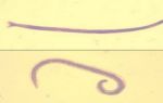
The length of the parasite is 1.2-4.5 mm. The genital openings, unlike echinococcus, are located in the anterior half of the hermaphrodite segment. The testes are concentrated in the posterior half of the proglotids. The uterus of mature segments has a sac-like shape, without lateral outgrowths characteristic of echinococcus. Oncospheres are the same as those of Echinococcus.
There are significant differences in the structure of finnose bladders (larvocysts) - they consist of many bubbles (brood capsules) filled with liquid or a jelly-like mass. Each vesicle contains 1-3 scolex. This multi-chamber structure is the result of the division of bubbles by ligation. On the surface of such a conglomerate of brood capsules there are small vesicles with scolex, which grow outward and grow into the surrounding tissues.
The diameter of alveococcal larvocysts in humans is from 2-5 to 10-15 cm. Occasionally they reach the size of an adult’s head.
Life cycle
A diagram of the alveococcus life cycle is presented here .
The final (definitive) hosts for alveococcus are wild carnivores:
- foxes,
- arctic foxes,
- wolves,
- jackals,
- dogs,
- rarely cats.
Intermediate hosts are mainly rodents (many species of voles, muskrats, and less commonly mice).
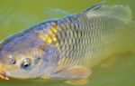
A person becomes infected accidentally, acting as an intermediate host - a biological dead end for alveococcus.
From eggs swallowed by a person, oncospheres emerge in the stomach, which penetrate the small intestine and from there through the portal vein system to the liver, where most of them are retained. Some part goes through the pulmonary circulation into the lungs, and the other part through the systemic circulation into many organs (bones, brain, spleen, etc.).
The most typical development of larvocysts (Finn) is in the liver, where degeneration of the hepatic parenchyma occurs around them.
As Finns grow, fibrous connective tissue forms around them, which becomes scar.
As a result, nodes are formed in the form of a whitish tumor with many cysts connected to each other. Due to the proliferation of vesicles located on the periphery of the finna, they actively grow into neighboring organs and tissues. This resembles the growth of a malignant tumor. In addition, individual vesicles break off from the surface of the node and larvocysts metastasize to other organs. When Finn is localized in the liver, blockage of the bile ducts occurs, accompanied by jaundice.