Alveococcosis is a biohelminthiasis caused by the larval stage of the tapeworm Echinococcus granulosus (alveococcus).
Read about alveococcus in the article “ Alveococcus - life cycle, structure .”
Content
Where can you get infected?
This is a natural focal disease. In Russia, foci of the disease exist in Kamchatka, Chukotka, Yakutia, Krasnoyarsk, Khabarovsk territories, and Western Siberia.
Abroad - in Switzerland, Austria, Germany, France, Bulgaria, Turkey, Japan. The incidence of alveococcus in different regions of Russia is 0.4-8.8 per 100 thousand population. Children get sick more often.
Routes of infection
Sources of infection are often wild animals - foxes, wolves, arctic foxes, and less often - domestic cats and dogs. Mature helminth eggs are shed in animal feces, contaminating the fur and the environment.
Human infection occurs through the mouth in 3 ways:
- directly from animals when they swallow oncospheres located on their fur (this is how hunters, members of their families, fur harvesters, fur farm workers become infected);
- when eating wild berries and herbs, drinking water from open natural sources and melt water;
- in close contact with infected dogs.
How does the disease develop?
In the human gastrointestinal tract, alveococcal oncospheres are released from the membrane, the released larvae penetrate into the mesenteric blood vessels and are carried by the bloodstream. Most of the larvae remain in the liver. Usually the right lobe of the liver is affected, where nodes with a diameter of 0.5-30 cm are formed, which grow into the bile ducts, diaphragm, and kidney. Dystrophy and atrophy occur in the affected organ. Compensation of liver functions occurs due to hypertrophy (enlargement) of healthy areas of the organ.
Obstructive jaundice appears, and in the later stages - biliary cirrhosis. Due to active infiltrative growth, a decay cavity is formed in the middle of the alveococcal node, which is often filled with purulent contents.
Symptoms
Alveococcosis most often affects young and middle-aged people (20-50 years old).
The incubation period is months, sometimes years.
Highlight
- alveococcosis of the liver and
- extrahepatic alveococcosis.
During the course of the disease there are 3 stages:
- early uncomplicated
- stage of complications
- terminal.
Early uncomplicated stage
In the early stages, the patient may not be aware of the infection. It is asymptomatic, although there are already small alveococcal nodes in the liver. During a routine examination, they can be determined even by palpation.
Some patients experience allergic reactions in the form of urticaria, sometimes with itchy skin.
As the node grows in the liver,
- heaviness and pain in the right hypochondrium and epigastrium,
- bitterness in the mouth,
- nausea,
- belching.
Gradually, pain in the liver area increases, attacks of biliary colic periodically occur, all the above symptoms increase, and upon palpation a low-painful “stone” liver is discovered.
Stage of complications
At this stage, obstructive jaundice, purulent cholangitis appear, and an abscess is possible.
When the alveococcus is compressed or grows into the gates of the liver, symptoms
- portal hypertension,
- ascites (fluid in the abdominal cavity),
- enlarged spleen,
- dilation of the vessels of the anterior abdominal wall,
- varicose veins of the esophagus and stomach.
With the formation of decay cavities in alveococcal nodes
- body temperature rises,
- pain intensifies,
- headaches appear
- general weakness.
Germination into the diaphragm and lung leads to a breakthrough of the contents of the decay cavity into the bronchi and the death of the patient from asphyxia or severe pneumonia. The rupture of the contents of the decay cavity into the abdominal cavity causes peritonitis, which is manifested by symptoms of an acute abdomen .
The most severe complications of the disease are associated with metastasis of the alveococcus to the brain and lungs, and less often to other organs.
When the brain is damaged, they appear
- Jacksonian seizures,
- monoparesis,
- sensory disturbance,
- headache,
- dizziness,
- nausea,
- vomit.
Metastases in the lungs appear
- chest pain
- cough with scanty mucopurulent sputum,
- detection of focal shadows on chest x-ray.
Allergic symptoms may also appear
- skin itching,
- hives,
- increase in eosinophils in the blood.
In the terminal stage of the disease, irreversible metabolic disorders develop.
Diagnostics
Differential diagnosis is carried out with various tumor-like formations of the liver:
- cystic echinococcosis,
- neoplasms,
- polycystic disease,
- hemangioma,
- cirrhosis.
When diagnosing alveococcosis, the same methods are used as for cystic echinococcosis .
A detailed survey of the patient (place of residence, profession and occupation, degree of contact with dogs and wild carnivores) plays an important role.
It is important to pay attention to clinical features:
- slow increase in symptoms
- long-term course, not amenable to conservative treatment,
- the presence of an allergic component (itching, rash).
In the blood test:
- variable eosinophilia (up to 15%),
- increase in ESR,
- hyperproteinemia (100-110 g/l),
- hypoalbuminemia,
- hypergammaglobulinemia,
- increase in thymol test, C-reactive protein.
Ultrasound , CT, NMR imaging, and angiography can determine the localization, extent of the process, and the presence of metastases
During X-ray examinations, cysts in the liver or lungs appear as round shadows with clear contours. Around cysts in the liver, rings of calcification in the form of lime splashes are often found.
Laparoscopy is rarely used.
Treatment
Treatment of alveococcosis is surgical. If surgery is not possible, chemotherapy is performed in combination with symptomatic therapy.
It is rarely possible to radically remove a parasitic node due to significant damage to the organ. When nodes grow into the porta hepatis, inferior vena cava, or neighboring organs, partial resection (removal) is performed, and the remaining parasite tissue is subjected to cryodestruction or treatment with chemical antiparasitic agents.
Chemotherapy is indicated for all patients, with the exception of the terminal stage, to prevent relapse of the disease. It is also justified in inoperable cases.
For the treatment of the disease, the drug of choice is albendazole .
It is prescribed internally:
- adults with a body weight of more than 60 kg - 400 mg 2 times a day with meals;
- with body weight less than 60 kg and for children - 15-20 mg/kg body weight in 2 divided doses (morning, evening).
The duration of treatment for solitary alveococcal liver nodule is at least 5 years without interruptions. In case of combined damage to several organs or the brain, treatment is carried out for life.
Further observation of patients
Dispensary observation after a course of treatment is lifelong, examination frequency is 1-2 times a year.
The following examinations are carried out:
- general blood and urine analysis;
- biochemical blood test - ALT, AST, bilirubin, prothrombin, proteinogram, serological reactions;
- chest x-ray;
- Ultrasound of the abdominal organs;
- CT.
If necessary, examination by a neurologist, gastroenterologist, or pulmonologist.
Prevention
The likelihood of infection is especially high when processing fur raw materials.
Measures should be taken to prevent human infection from dogs and fur-bearing animals kept in an enclosure.
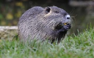
To reduce the likelihood of infection of dogs and cats, it is necessary to eliminate the possibility of them feeding on rodents: they should not be fed the carcasses of mouse-like rodents, muskrats, hamsters, beavers and other rodents - possible intermediate hosts of alveococcus.
To avoid infection, you should follow the rules of hygiene when hunting and fishing, and do not eat unwashed wild berries and herbs.