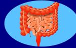
An abdominal hernia is the emergence of internal organs from the abdominal cavity under the skin along with the peritoneum lining the abdominal cavity through natural or artificially formed holes in the wall of this cavity.
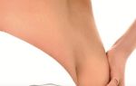
Umbilical hernia is a condition in which the internal organs (intestines, greater omentum) extend beyond the anterior abdominal wall through an opening located in the navel area.
Content
- 1 Description
- 2 Reasons for education
- 3 Types of hernias
- 4 Hernias in men
- 5 Hernias in women
- 6 How to recognize an umbilical hernia yourself?
- 7 Common symptoms of abdominal hernias
- 8 Complication
- 9 Symptoms of strangulation
- 10 Diagnostics
- 11 Treatment
- 12 Types of operations
- 13 Rehabilitation after surgery
- 14 Prevention
Description
Hernial protrusion consists of
- hernial sac (peritoneum),
- hernial ring or gate,
- hernia contents.
The hernial orifice (ring) is an opening in the muscle-tendon part of the abdominal wall.
The contents are most often the omentum or intestinal loops.
If the contents of the hernia are freely reduced into the abdominal cavity when lying down or pressing on the hernial sac, it is called reducible.
If adhesions develop between the contents of the hernia and the hernial sac or the contents are so large that they do not go into the abdominal cavity, the hernia becomes irreducible.
Reasons for education
The reasons are divided into two groups -
- factors that predispose to the formation of a hernia and
- factors that directly cause the formation of a hernia.
Predisposing reasons are
- poor development of muscles in certain areas of the abdominal wall,
- weakening of the abdominal wall due to sudden weight loss,
- with significant obesity , muscle tissue is replaced by fatty tissue,
- genetically determined or age-related thinning,
- loss of tissue elasticity.
A hernia may form
- when intra-abdominal pressure increases, for example, when lifting heavy weights that do not correspond to the physical development of a given person;
- with repeated pregnancies;
- constipation;
- chronic cough;
- crying in infants;
- difficulty urinating (phimosis in children, narrowing of the urethra in old people, etc.).
The occurrence of an abdominal hernia is due to the anatomical structure of the abdominal wall and the presence of so-called weak spots in certain places. Increased pressure in the abdominal cavity leads in these weak areas of the abdominal wall to the divergence of muscle fibers and the formation of a gap through which the peritoneum protrudes; The abdominal organs fall into the peritoneal sac thus formed.
Types of hernias
According to the place of origin they distinguish
- inguinal,
- femoral,
- umbilical,
- internal and
- other hernias.
The most common are inguinal, in which the insides, most often the intestines, exit the abdominal cavity through the inguinal canal. The inguinal canal is a narrow slit-like space located obliquely (from top to outside, inside and down) in the lowest part of the anterior abdominal wall, in the groin area.
Hernias in men
In a male fetus, the testicles descend from the abdominal cavity into the scrotum through the inguinal canal. In adult men, the spermatic cord passes through the inguinal canal, and in women, the round ligament of the uterus.
Inguinal hernias in boys can be congenital due to improper development of the inguinal canal and delayed descent of the testicles into the scrotum.
Hernias in women
In femoral hernias, which most often develop in women, the viscera exits the abdominal cavity through the femoral canal. The femoral canal, as such, does not exist. This name refers to the place where large vessels of the lower limb exit from the abdominal cavity onto the thigh.
On the side of the abdominal cavity in this place there is a depression - a fossa. Under unfavorable conditions, protrusion of the abdominal wall occurs in this area, followed by the formation of a hernial sac and the development of a hernia.
Children and women often develop hernias in the navel area, so-called umbilical hernias, and in men, hernias of the white (middle) line of the abdomen.
A special place is occupied by hernias that develop in the area of extensive scars that occur in places of injury to the abdominal wall or in the area of postoperative scars - postoperative hernias - if the surgical wound took a long time to heal with symptoms of suppuration.
How to recognize an umbilical hernia yourself?
An umbilical hernia can be recognized independently. The main symptoms of an umbilical hernia are:
- the appearance of a protrusion in the navel area,
- sharp pain in the navel area when coughing or moving,
- reduction of pain and protrusion when lying on your back.
If you recognize symptoms similar to an umbilical hernia, we recommend that you consult a surgeon for an accurate diagnosis and subsequent treatment. An umbilical hernia without treatment tends to increase in size, which can lead to complications and require more serious intervention.
Common symptoms of abdominal hernias
The main sign of a hernia is the appearance of a protrusion in a typical location for a hernia (groin area, upper thigh, umbilical area, scar after surgery, etc.). The protrusion increases with tension and disappears when lying down.
There may be no pain or nagging pain when standing and working, which disappears when lying down.
The protrusion can exist for many years without significant increase, but more often there is a gradual increase in the size of the hernia, and the contents cease to be reduced even when lying down.
During reduction, the hernial ring is felt through the skin - that opening in the wall through which the viscera protrudes.
Complication
The retention and accumulation of intestinal contents in the intestinal loop trapped in the hernial sac causes a complication called coprostasis.
The most severe and dangerous complication is a strangulated hernia, which can lead to death if surgery is not performed in a timely manner. Incarceration often develops during muscle tension, for example, when lifting heavy objects, during bowel movements, coughing, etc. In these cases, when the pressure in the abdominal cavity increases, intestinal loops slip into the hernial sac, which are strangulated in the hernial orifice. In this case, compression of the vessels feeding the intestinal wall occurs; hemorrhages appear in the intestinal wall, its swelling, followed by necrosis of part of the wall, and sometimes the entire intestinal loop. Bacteria penetrate into the abdominal cavity through the dead intestinal wall and inflammation of the peritoneum develops - peritonitis.
Compression of the intestinal wall in the hernial orifice can cause the cessation of passage of its contents through the intestines, as a result of which intoxication (poisoning) occurs with products absorbed from the intestines, and develops intestinal obstruction.
Symptoms of strangulation
The main signs of a strangulated hernia are
- the appearance of pain in the area of the hernial sac,
- irreducibility of a previously repaired hernia,
- hernia enlargement,
- tension,
- pain when palpating the hernia, especially in the hilum area,
- nausea,
- vomit,
- lack of stool
- bloating,
- and with increasing intoxication - increased heart rate, general weakness, headache.
Diagnostics
- The doctor can determine the presence of a hernia during examination. He palpates the abdomen and asks the patient to cough to see changes characteristic of a hernia.
- Using ultrasound , the size of the hernia contents, the presence of any organ in the hernial sac, the presence of compaction and fusion of connective tissue are determined.
- To determine which organ is in the hernial sac, herniography is performed. Under anesthesia, a contrast solution is injected into the patient with a needle, and he is asked to turn over on his stomach and strain or cough so that the solution penetrates into the hernial sac. After this, x-rays are taken.
- Instead of herniography, it is possible to perform radiography using contrast fluid.
- In rare cases, the doctor also prescribes a computed tomography scan and endoscopic examination of the stomach.
Treatment
The main method of treating a strangulated hernia is emergency surgery. If it is done quickly, in the first hours after strangulation, before the development of necrosis of the intestine, it is not severe.
When the intestines become necrotic, the disease becomes very dangerous. In this case, part of the intestine is removed during surgery.
Any attempts to reduce a strangulated hernia are unacceptable, since this may damage the strangulated organ, or an inflamed or dead intestinal loop may be inserted into the abdominal cavity, which leads to severe peritonitis.
Sometimes a strangulated hernia can repair itself. In this case, the patient should see a doctor as soon as possible after the reduction for a thorough examination, and should undergo surgery at the first opportunity.
To avoid complications, any hernia is subject to surgery, even if it does not cause any disorders. Only for umbilical hernias in young children is non-operative treatment possible by applying adhesive bandages after reduction.
Retaining the hernia with a bandage does not protect against strangulation. The bandage causes weakening of the muscles, expansion of the hernial ring, and consequently an increase in the hernia. It is acceptable to wear a bandage for some time if surgery is contraindicated for health reasons.
Types of operations
An operation to remove a hernia using one’s own tissue is performed for small hernias (3 - 5 cm).
The most effective method is suturing the hernial orifice using local tissue. During the operation, minimal tissue dissection is performed and the hernia opening is tightened with stitches.
After this, the incision is closed and the suture is repaired, after which there are practically no visible traces of the operation. If there is a small layer of subcutaneous fat, surgery can be done using a surgical laser. The operation to remove a small hernia in adults is minimally invasive and does not require hospitalization; the complete recovery process does not exceed one month.
Relapses after a well-performed operation are also excluded. An operation to remove a hernia using a mesh implant is used
when the hernia is of a significant size, since tightening the hernial orifice with one’s own tissues is no longer a complete guarantee of recovery or requires serious surgical intervention. In this case, the method of implanting a polymer mesh implant is used. An obstructive mesh is applied to the hernial orifice, which is sewn with stitches to the surrounding tissues of the abdominal cavity. The mesh is made of a special material that is not rejected by the body and integrates well with the tissue of the abdominal wall. Within two to three weeks, the implant grows into the surrounding tissue and completely covers the hernial orifice with a dense connecting septum.
Surgery to treat an umbilical hernia using an implant can also be performed on an outpatient basis and does not require hospitalization. The implant can be introduced through a minimal tissue incision using the laparoscopic method, which eliminates subsequent scars on the skin. One month after the operation, all motor restrictions are removed. Relapses and complications with this method are practically excluded.
Rehabilitation after surgery
After hernia repair surgery (hernioplasty), the patient must undergo a rehabilitation course. In the first two weeks, it is recommended to wear a bandage and complete absence of physical activity. In the third week after surgery, if there is no pain, you can begin to do small exercises to strengthen the abdominal walls. Full recovery occurs a month after the operation, after which you need to move on to preventive procedures: strengthening the abdominal muscles, losing weight and strengthening the overall tone of the body.
Prevention
The main method of preventing the development of a hernia is physical exercise.
Wearing a bandage during pregnancy and abdominal massage after childbirth is of great importance for preventing hernia in women.
Should be eliminated with appropriate treatment
- constipation,
- cough,
- difficulty urinating, etc.
It is necessary to avoid obesity and sudden weight loss.
When caring for a child, everything that increases his intrauterine pressure should be eliminated:
- tight swaddling,
- early transfer of the child to a vertical position,
- tossing, etc.
Good and proper nutrition of a child strengthens his muscular system,
Proper ligation of the navel and careful care of the navel of a newborn is the prevention of umbilical hernias.