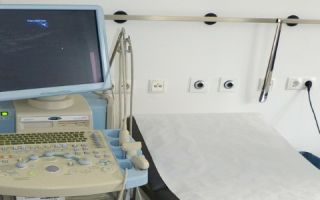
Ultrasound is the recognition of pathological changes in organs and tissues of the body using ultrasound. The method is based on the principle of echolocation - the reception of signals sent and then reflected from the surface at the interface of different media with different acoustic properties; it is a non-ionizing research method.
Content
How is ultrasound performed?
An ultrasound examination of the abdominal organs usually lasts 20–30 minutes. During the procedure, the patient must be in a supine position. The doctor applies a special transparent gel to the skin, places an ultrasound probe on the area being examined and slowly moves it. During the procedure, the patient does not experience any discomfort.
Do I need to prepare for an ultrasound?
3 days before the test, it is recommended to exclude gas-forming foods (milk, brown bread, greens, legumes, apples, cabbage, grapes, etc.). It is advisable to take activated carbon 2 tablets 3 times a day.
The study is carried out strictly on an empty stomach, preferably in the morning. Before the test, you should not eat, drink, chew gum, suck hard candies, smoke, or take medications. If the study is planned for the second half of the day, 7 hours before the study a light breakfast (tea, kefir, bun) is allowed, followed by a period of fasting - do not eat or drink anything.
To determine the contractile function of the gallbladder, have 2 bananas or a 200 ml package of cream with at least 10% fat or 2-3 grams of butter on a small piece of white bread.
No preparation is required to examine infants.
For patients who suffer from diabetes, a small breakfast (warm tea, bread) is allowed before the study of the kidneys and liver. You should not smoke before the ultrasound procedure, as this causes contractions of the stomach, which may lead to the doctor making an incorrect diagnosis. If you use any medications, you need to warn your doctor about it.
What does an abdominal ultrasound show?
Using ultrasound, various volumetric formations of both internal organs and superficial tissues (cysts, tumors) are detected with fairly high accuracy.
In severe forms of the disease, diagnosis by ultrasound examination is quite accurate. In the early stages of the disease, only changes in organs characteristic of a particular diagnosis can be detected.
Ultrasound of the intestines
During a screening examination of the intestines, an ultrasound examination allows you to determine the presence of diseases such as:
- tumors of the small and large intestine,
- intestinal tuberculosis,
- intestinal diverticulosis,
- Crohn's disease,
- ulcerative colitis,
- acute appendicitis,
- intussusception and mechanical intestinal obstruction.
Once the diagnosis is established, ultrasound can be used to examine the condition of the intestinal wall.
The thickness of the walls of the small and large intestines in an ultrasound image is normally 2-6 mm. The maximum diameter of the small intestine does not exceed 40 mm, and the maximum diameter of the large intestine is 60 mm.
These proportions change with thickening of the intestinal wall due to edema, fibrosis, hemorrhage, tumor damage, or the transfer of the inflammatory process from neighboring organs. In this case, the peripheral ring expands, and the central part looks relatively small. This sign has different names among specialists: “pseudo-kidney”, “target”, “bull’s eye” or “symptom of an affected hollow organ”.
With ultrasound, you can sometimes observe a pendulum-like movement of the intestinal contents.
Advantages of ultrasound when examining the intestines
Unlike X-ray and endoscopic examinations, ultrasound makes it possible to evaluate the entire intestinal wall down to the serous membrane, its external contours and neighboring organs.
With the help of repeated ultrasound examinations, it is possible to track the dynamics of the disease in patients with ulcerative colitis, Crohn's disease, and intestinal tuberculosis and diagnose complications.
For early diagnosis of colon tumors, endoscopic ultrasound of the colon and rectum is used.
Ultrasound of the liver
When examining the liver, you can reveal:
- cirrhosis of the liver,
- ascites (fluid in the abdominal cavity),
- increased diameter of the portal vein and spleen,
- cysts,
- fatty hepatosis.
Various changes in the liver allow us to draw a conclusion about a particular disease. First of all, attention is paid to such anatomical changes as:
- tissue swelling,
- fatty infiltration,
- sclerosis of the walls of the hepatic arteries,
- varicose veins,
- tissue fibrosis.
Depending on the severity of certain signs and their combination, a diagnosis is made.
Signs of acute hepatitis on ultrasound
- A uniform increase and significant decrease in the echogenicity of the liver parenchyma.
- Expansion of the portal vein and its segmental branches.
- Increased echogenicity of tissue along the gallbladder.
- In 30% of cases, an enlargement of the spleen and gall bladder is observed.
- Enlargement of the pancreas and decreased echogenicity of its parenchyma.
Signs of liver cirrhosis
- Diffuse or focal heterogeneity of the liver structure.
- Many obliterating vessels.
- Enlargement of one lobe of the liver with atrophy of the other.
- Rounding of the lateral segment.
- Ascites (fluid in the abdominal cavity).
- Dilatation of the portal vein.
- Enlarged spleen (splenomegaly).
- Gallbladder with signs of cholecystitis.
Signs of chronic hepatitis
- Enlargement of all lobes of the liver.
- Diffuse and uneven echogenicity of the image.
- Multiple obliteration of blood vessels (fusion of the lumen).
- Tortuous dilated veins.
- The spleen and pancreas are unchanged.
Gallbladder examination
The gallbladder normally has an elongated shape, dimensions within 10x4 cm, wall thickness not exceeding 0.4 cm.
Ultrasound of the gallbladder allows you to diagnose:
- congenital anomalies (double gallbladder, diverticulum, presence of septum, etc.),
- tumors and cholesterol polyps,
- concretions (stones),
- inflammatory changes (manifested by wall thickening of more than 0.4 cm).
Ultrasound allows you to most accurately determine changes in the gallbladder. If chronic and calculous cholecystitis is suspected, the final diagnosis is made by ultrasound examination.
A healthy gallbladder has an oblong shape with a clear echogenic cavity and thin walls.
Signs of changes in the gallbladder are:
- wall thickening,
- deformations, deformations
- the presence of partitions in the cavity,
- heterogeneity of echogenicity of the cavity,
- the presence of individual shapeless foci of echogenicity in the parenchyma surrounding the gallbladder,
- reduction in the size of the gallbladder,
- increase in the size of the gallbladder.
Only three of these seven signs (deformation, presence of partitions and changes in size) are detected by radiography.
Signs of chronic cholecystitis on ultrasound
- Thickening of the gallbladder wall (especially evident on an empty stomach).
- Deformation of the gallbladder is a violation of the normal oval shape of the organ, shapeless contour outlines.
- Scar changes in the cervical area.
- The presence of septa, which are visualization of individual scars and adhesions.
- Fibrous changes in the parenchyma surrounding the gallbladder.
- Heterogeneity in the image of the gallbladder cavity is a sign of stones or papillomas. The image of stones is easily diagnosed by the presence of a “shadow path” behind them. The papilloma does not shift when the patient’s body position changes.
- An increase in the size of the gallbladder indicates a decrease in excretory function as a result of cicatricial changes or partial obstruction due to inflammation of the major duodenal papilla.
- A reduction in the size of the gallbladder may be the result of scar changes due to chronic cholecystitis or congenital hypoplasia.
Signs of blocked bile ducts
Undilated bile ducts have a diameter of 1-2 mm and are normally not visible. The diameter of the common bile duct is an important indicator of bile duct obstruction, even more important than the diameter of the intrahepatic bile ducts.
Normally, the diameter of the common bile duct is 4-5 mm. A diameter of 6 mm indicates dilation of the bile ducts.
The diameter of the extrahepatic bile ducts increases with age and in patients who have undergone surgery to remove the gallbladder.
Therefore, their increase is not always a sign of blockage. An accurate diagnosis can be made by repeat scanning after ingesting fatty meat foods or internal administration of cholecystokinin. If the diameter of the duct does not change in size after repeated scanning, then there is a blockage of the duct.
Sonography
This ultrasound examination method is the most reliable method for diagnosing subhepatic jaundice. In this case, signs of jaundice are dilation of the bile ducts and gallbladder. These data make it possible to distinguish subhepatic jaundice from hepatic jaundice, in which dilatation of the bile ducts is not observed.
Pancreas
Ultrasound can detect acute and chronic pancreatitis.
Acute pancreatitis is characterized by:
- enlarged pancreas;
- poor visibility of the splenic and portal veins.
- Signs of chronic pancreatitis are:
- enlarged pancreas;
- unevenness, sometimes blurriness, of contours;
- dilation of the pancreatic duct, which is not normally visible;
- formation of pseudocysts.
Ultrasound of the spleen
During the examination, the size of the spleen is assessed, which normally should have a crescent shape. This study for splenomegaly (pathological enlargement of the spleen) allows us to determine the reasons for the enlargement of the organ - tumors, cysts, hematomas.
The condition of the spleen is also important to evaluate in case of liver diseases. With cirrhosis of the liver, there is an enlargement of the spleen and the presence in its parenchyma of obliterated vessels (with closure of the lumen), which are absent in hepatitis.
The width of the splenic vein is also an important indicator.
In difficult diagnostic situations, a highly informative but unsafe method is used - laparoscopy.


