Paragonimiasis is a biohelminthiasis that is caused by a pulmonary fluke and most often affects the lungs, less often the brain and other organs.
Content
All about the pulmonary fluke
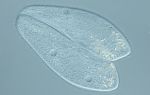
This parasite belongs to trematodes, flatworms. Sexually mature individuals (maritas) are ovoid in shape, 7.5-16 mm long, and the body is covered with spines. They parasitize the lungs of humans and a number of carnivorous animals (dogs, cats, pigs). This is where the flukes lay their eggs. Through the bloodstream, the egg enters various organs of the final host, sometimes the brain, causing organ damage and inflammation.
The eggs leave the human body with sputum, and when the sputum is swallowed, the eggs are found in the stool.
The eggs of the pulmonary fluke are quite large - 60-100 microns, golden brown in color, with a cap. When released into freshwater bodies, miracidium emerges from the eggs and actively penetrates the body of mollusks of the genus Melania. In the body of the first intermediate host, the parasite passes through the stages of sporocyst, redia and circaria. The latter leave the body of the mollusk and actively invade crayfish and crabs (second intermediate hosts).
Humans and animals are the definitive hosts for these worms and become infected by eating undercooked crayfish and crabs.
What is paragonimiasis?
This is biohelminthiasis, which is caused by a pulmonary fluke and most often affects the lungs, less often the brain and other organs.
The disease is common in Central and East Asia (China, Taiwan), East Asia (DPRK, South Korea, Japan), Southeast Asia (Vietnam, Indonesia, New Guinea, Solomon Islands, Laos, Thailand, Philippines) , much less, in the form of individual foci, in South Asia (India, Sri Lanka). In Russia, in addition to imported cases, limited foci of paragonimiasis are known in the Primorsky Territory and in the Amur region.
The definitive hosts of the parasite - pigs, dogs, cats and wild carnivores and humans - excrete helminth eggs in sputum and feces. Intermediate hosts are mollusks, additional hosts are freshwater crayfish and crabs.
How is it transmitted?
Pulmonary fluke can be contracted through food and water. Poorly cooked freshwater crayfish and crabs are contaminated foods.
When crayfish and crabs die, the parasite leaves their bodies and remains alive in the water for 25 days. Therefore, infection is possible by swimming in water bodies, as well as by drinking contaminated water.
Development of the disease
After eating contaminated meat of crayfish and freshwater crabs in the duodenum, the larvae are released from the membranes and perforate its wall, entering the abdominal cavity. Then it rises up, drills through the diaphragm and penetrates into the lungs.
In the development of paragonimiasis, an important role is played by mechanical damage to body tissues by worms and their eggs, as well as toxic-allergic reactions of the body to the waste products of the parasite.
During the migration of parasite larvae into the lungs through the diaphragm and other organs (liver, pancreas, kidneys), hemorrhages and sometimes necrosis occur. In the lungs (especially in the lower lobes), in addition to hemorrhages, eosinophilic infiltrates are formed.
From about the second month, fibrous cysts with a diameter of 0.1-10 cm are formed around the parasite near the roots of the lungs and along the periphery of the lung tissue. A human cyst usually contains one, rarely two, parasites. They reach sexual maturity and begin laying eggs 5-6 weeks after infection. Cysts are often connected to the branches of the bronchi. After the death of the pulmonary fluke or its release from the cyst, the cavity becomes scarred and petrified.
If the wall of the cyst is broken, parasites or their eggs enter the blood and are carried into the brain, mesentral lymph nodes, prostate gland, liver, skin and other organs and tissues.
Symptoms
The incubation period lasts 2-3 weeks, with massive infection it is reduced to several days.
In the acute phase, paragonimiasis occurs as an acute allergic disease with severe allergy symptoms:
- skin rash and itching;
- redness of the mucous membranes;
- runny nose, cough.
Found in the lungs
- eosinophilic infiltrates,
- pneumonia,
- pleurisy.
There are two stages in the acute phase
- acute abdominal and
- acute pulmonary-prevular paragonimiasis.
In the first stage, symptoms develop
- severe enteritis,
- hepatitis A,
- benign aseptic peritonitis with symptoms of acute abdomen .
At the second stage
- body temperature rises,
- there are chest pains,
- dyspnea,
- cough with purulent sputum,
- sometimes hemoptysis.
Physical examination and x-ray examination reveal infiltrates.
After 2-3 months, the disease enters the chronic stage with remissions and exacerbations.
During exacerbations
- body temperature rises to 38-40 degrees,
- chest pain intensifies,
- headaches appear
- dyspnea,
- cough, with the release of rusty sputum, which contains helminth eggs,
- Hemoptysis is often noted.
After 2-4 years, the clinical manifestations of the disease gradually disappear. After the disappearance of the symptoms of the disease, an X-ray examination of the lungs reveals small isolated foci of fibrosis and single or multiple calcifications with a diameter of 2-5 mm.
With intensive infection and a long-term course of the disease, pneumosclerosis and pulmonary heart syndrome develop.
The entry of worms into the central nervous system causes
- meningitis,
- increased intracranial pressure,
- possible atrophy of the optic nerve,
- paresis,
- paralysis,
- sensory disturbances,
- epilepsy.
X-rays of the brain in such patients reveal calcified round formations containing dead helminths.
Diagnostics
When diagnosing, pneumonia, tuberculosis, and tumors are excluded.
The parasitic form of the disease is indicated by the combination of neurological symptoms with characteristic changes in the lungs and the presence of parasite eggs in sputum and feces.
In the early period, when young parasites have not yet released eggs, ELISA is used for diagnosis (test systems are not registered in the Russian Federation). You can use an intradermal allergy test with antigens from Paragonimus.
In endemic areas, residents of settlements located along the banks of rivers, lakes, and reservoirs (at least 20% of residents once every 2-3 years), workers of fish processing plants, fishing boats and their families are examined annually.
Drug treatment
Treatment should be carried out after relief of allergic phenomena.
The drug of choice is praziquantel . It is prescribed to adults and children over 4 years of age at a daily dose of 75 mg per 1 kg of body weight in 3 doses with an interval of 4-5 hours for 2 days.
Alternative drugs are triclabendazole, bithionol, which are taken as for fascioliasis .
In case of damage to the central nervous system, specific treatment is carried out in a hospital due to the possible development of cerebral edema and increased intracranial pressure. In this case, diuretics and anticonvulsants are used. Single cysts with helminths are removed surgically.
To monitor the effectiveness of treatment, 1 month after therapy, once with a persistent cough, sputum is examined for pulmonary fluke eggs.
Prevention and prognosis
The prognosis for modern treatment of uncomplicated cases is favorable. With a widespread process, the prognosis is serious.
To prevent infection, you should
- follow technological methods for preparing dishes from freshwater crayfish and crabs,
- prevent fecal pollution of water bodies,
- Use only boiled or filtered water for drinking.