Amebiasis is a disease caused by dysenteric amoeba, which leads to ulcerative lesions of the colon with abscesses in the liver, brain, lungs and other organs.
Content
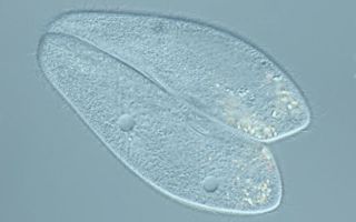
Amoeba
Several species of amoebas live in the human digestive system. The harmful effects on the body of some of them have not been proven.
Oral amoeba
It is found in 20-25% of people during examination. The oral amoeba enters the human body through airborne droplets with droplets of saliva or sputum when an infected person sneezes and coughs, when using the same toothbrush, eating utensils, or when kissing.
In the oral cavity, amoebas live between teeth, in gum pockets, and in tooth cavities formed as a result of caries.
They may be the reason
- bad breath
- increased formation of tartar,
- complications with sinusitis, pneumonia, osteomyelitis.
Intestinal amoeba
Intestinal amoeba (Entamoeba coli) is found quite often both in healthy people and in patients with intestinal diseases. Its effect on human health has not been proven.
Dysenteric amoeba
It is the dysenteric amoeba (Entamoeba histolitica) that is the causative agent of amoebiasis. This type of amoeba is common on all continents, but the disease is more common in countries with tropical and subtropical climates.
Once in the human intestine, amoebas develop and divide. They feed on colon bacteria. As they move through the intestines, they turn into cysts and pass out with feces.
When the body's defenses are weakened due to
- hypothermia,
- stress, stress
- lack of vitamins,
- chronic diseases
- some forms of amoebas transform into hematophages. They begin to absorb red blood cells. After some time, an intestinal ulcer forms and the amoeba penetrates the blood vessels. It spreads through the blood throughout the body, reaching the liver, lungs, brain and other organs, forming abscesses in them.
Amoebiasis
In Russia, cases of amoebiasis are rare. These are mainly people who come from countries where this infection is widespread. These are the countries of Central Asia, Southeast Asia, Africa, South and Central America.
Some regions of the Russian Federation are also at risk: the south of Primorsky Krai, Dagestan. The disease also occurs in Georgia and Armenia.
How can you get infected?
Cyst forms of dysenteric amoeba secreted by sick people live in feces for up to 30 days, in the water of natural reservoirs from 9 to 60 days.
You can become infected with amoebas
- through the soil
- wastewater,
- water from open reservoirs,
- household items,
- fruits, vegetables, food products,
- dirty hands.
Types of amoebiasis
In accordance with WHO recommendations, there are:
- invasive amebiasis (penetration of amoebas into the intestinal mucosa and other organs),
- asymptomatic carriage.
According to the course of the disease, they are distinguished:
- extraintestinal amebiasis,
- intestinal amebiasis (amebic dysentery).
Amoebic dysentery is of the following types:
- asymptomatic infection
- amoebic dysentery,
- fulminant colitis with intestinal perforation,
- toxic megacolon,
- chronic amoebic colitis,
- ameboma,
- perianal ulceration.
Acute intestinal amebiasis (amebic dysentery)
After infection, it can take from several days to several months for the first symptoms to appear.
The development of the disease is characterized by symptoms such as
- bloating,
- pain in the right iliac region,
- copious, mushy stools, 3-5 times a day,
- a small amount of mucus and blood in the stool.
After some time, the stool becomes liquid with a large amount of glassy mucus, which is saturated with blood and looks like raspberry jelly. The frequency of stools is up to 15-20 times a day.
Sometimes the patient develops a painful false urge to defecate, burning and pain in the rectum, which intensifies during defecation.
In weakened people, body temperature rises to 39⁰C.
Chronic amebiasis (amebic dysentery)
Sometimes the acute period of amebiasis disappears, lasts 4-6 weeks and without specific treatment becomes chronic. For many years, periods of calm alternate with exacerbations. Chronic amoebic dysentery can last up to 10 years without treatment.
During exacerbations, constipation alternates with diarrhea, pain in the lower right or left half of the abdomen is bothersome. The state of health is not significantly affected; the body temperature remains normal.
During periods of remission, patients experience only minor intestinal disorders: flatulence, rumbling in the stomach, etc.
In this case, the ulcerative process gradually spreads to the entire colon. Against this background, anemia, hypopolyavitaminosis develop, and digestive functions are disrupted.
Complications
If left untreated, amoebic dysentery can lead to serious complications such as:
- intestinal perforation,
- peritonitis,
- amoebic appendicitis,
- intestinal obstruction,
- ameboma,
- amoebic lesion of the skin around the anus.
Perforation of the intestinal wall in amoebic dysentery
This complication is the most dangerous and is manifested by the following symptoms:
- acute pain in the abdomen,
- fever,
- abdominal muscle tension,
- other symptoms characteristic of an “ acute abdomen ”.
This complication most often develops at the height of the disease and can cause death.
In some cases, adhesive fibrous peritonitis develops.
This complication is manifested by the following symptoms:
- painful infiltrate 3-15 cm,
- increase in body temperature,
- local tension in the muscles of the anterior abdominal wall.
This type of peritonitis does not require surgery and can be easily treated with antiparasitic drugs.
Amoebic appendicitis
Amoebic appendicitis develops as a result of the spread of amoebae into the vermiform appendix (appendix). The complication is accompanied by signs of acute and chronic appendicitis .
The preferred treatment is with antiparasitic drugs, since surgery can cause amoebas to enter the bloodstream and spread these protozoa throughout the body.
Intestinal obstruction caused by amoebic dysentery
With a long course of amoebic dysentery, multiple ulcers of the colon become scarred and eventually intestinal obstruction with typical symptoms may develop.
Ameboma (amebic tumor)
If the disease continues for a long time without treatment, an amoebic tumor may form. Most often, ameboma forms in the cecum or ascending colon , less often in the hepatic or splenic flexures of the colon.
Surgical treatment is not required, since ameboma responds well to treatment with anti-amoebic medications.
Amebic skin lesions of the perianal area
In weakened individuals, the process extends to external tissues. Erosions and ulcers are located around the anus, in the perineum and on the buttocks.
Extraintestinal amoebiasis
In approximately 10% of all cases of disease, amoebas enter the bloodstream through colon ulcers and spread throughout the body.
When they enter the liver, they form abscesses in the right lobe, near the diaphragm or in the lower part of the liver.
The size of abscesses varies from barely visible to the eye to 10 cm or more.
Amoebas with blood can enter the lungs, brain, and genitourinary system.
Diagnostics
The diagnosis of amoebic dysentery is established only when pathogenic forms of dysenteric amoeba (hematophages) are detected in the patient’s feces.
For analysis, fresh material is used, which is collected no later than 15-20 minutes later. The material should not be stored in the refrigerator, since at low temperatures amoebas sharply lose mobility and are difficult to distinguish from other cellular elements.
If all signs of the disease are present, but hematophages are absent in the tests, the study is repeated 5-6 times a day. To stimulate the secretion of amoebas, a saline laxative is sometimes prescribed.
Instrumental studies are used to establish a diagnosis.
Colonoscopy and sigmoidoscopy ulcers of the intestinal mucosa with characteristic edges.
During endoscopy, material is taken for parasitological examination.
With radiography , CT and ultrasound , abscesses are identified, their location and size are determined.
The serological method allows you to identify antibodies produced by the body to dysenteric amoeba.
Treatment
Since serious complications can develop with amebiasis, it is recommended to treat it in a hospital.
To treat intestinal amebiasis (amebic dysentery), drugs such as:
- metronidazole,
- tinidazole,
- ornidazole,
- Secnidazole
When treating amoebic liver abscess, use:
- metronidazole,
- tinidazole,
- Secnidazole
Aspiration (percutaneous drainage)
This procedure is recommended for the following conditions:
- abscess size is more than 6 cm,
- localization in the left lobe of the liver or high in the right lobe of the liver,
- severe abdominal pain,
- tension in the abdominal wall due to the threat of abscess rupture,
- lack of effect from drug therapy.
Forecast
With early diagnosis and timely treatment, complete recovery is possible. With treatment, improvement in well-being occurs within a few days. Without treatment, the mortality rate for amoebic dysentery is 5-10%, and for extraintestinal complications - 50%.
Prevention
- Prevention measures are the same as for intestinal infections.
- After the course of treatment, you must be observed by a doctor for 1 year, taking tests once every 3 months.
- Compliance with hygiene rules.
- Drinking clean water.
- Washing fruits and vegetables with safe water.
- After traveling to epidemiologically dangerous countries, it is recommended to undergo testing for the early detection of intestinal ulcers and hidden infections.
- Immediately consult a doctor if you feel unwell after such travel.
- Disinfection of patient secretions and linen, routine disinfection in hazardous areas with a 3% Lysol solution and a 2% cresol solution.